musli :

Morphology:
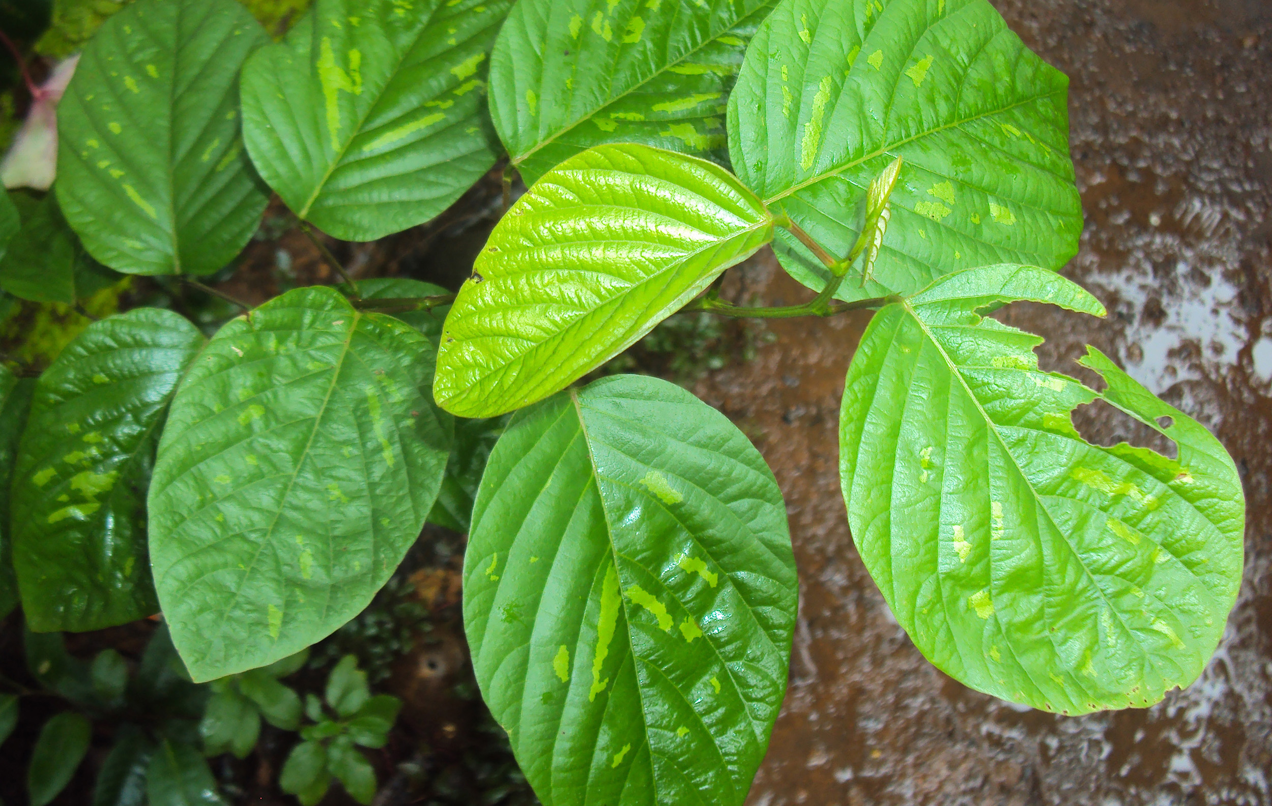
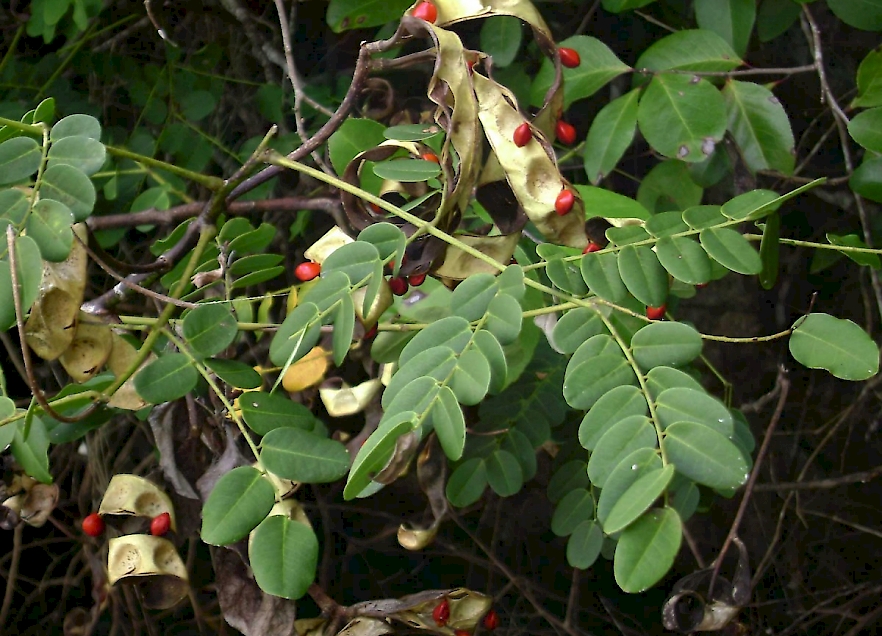
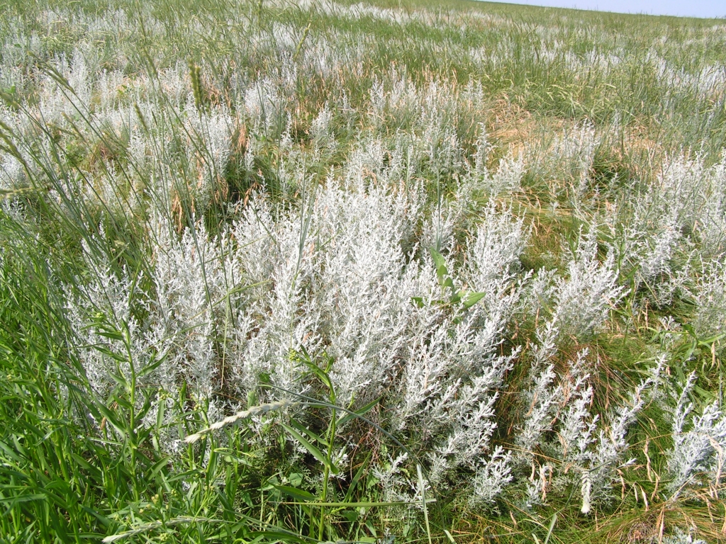
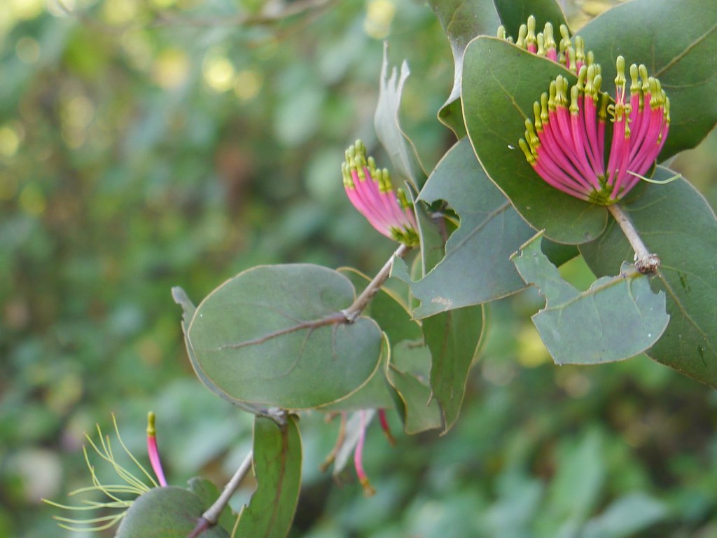
The plant form of Asparagus adscendens is a shrub of struggling nature much branched, spines with woody stem, It can grow up to an utmost height of 1.5 feet. Cladodes are 0.6-1.2 cm long linear in shape but stout, straight, bear spines . Flowers are small, white, 3-4 cm across, solitary or fascicled with copious racemes. Fruits are 0.8 cm in diameter, globes, 3 lobed berries with only one seed . Tubers can grow up to a depth of 10 inchHistology:
-T.S. of young stem showed presence of ridges and furrows along with most of the typical monocot characters such as large pith and irregularly arranged vascular bundles (Fig 2). Epidermis consisted of single layer of dumbbell shaped cells possessing thick layer of cuticle followed by cortex region. Cortex was seen differentiated into outer 3-4 layers of collenchymatous cells below ridges and furrows and an inner continuous layer of parenchymatous cells. Vascular bundles were found scattered irregularly with larger ones placed towards the inner side and smaller ones developing in peripheral region. Vascular bundles were of collateral and closed type with metaxylem present on its outer side. In addition centrally placed pith composed of larger cell size
2 Microscopic characters of cladodes
Horizontal section of cladodes viewed triangular in outline with a single cell layer of dumbbell shaped epidermal cells. Below the epidermis 2-3 palisade cell layers with centrally placed vascular tissue zone was present. Xylem and phloem were found intermixed in the central zone.
3 Microscopic characters of the root Transverse section of root clearly showed the outermost layer of epidermal cells, compactly arranged, thick walled cells forming the piliferous layer. Below the epidermis two types of cortex were present i.e. outer lignified cortex and inner parenchymatous cortex. Sclerenchymatous fibres were found scattered in the cortex while some of them on maceration appeared as scattered fibres. A well-developed sheath of stone cells surrounding the endodermis was present at all levels of root. The innermost 1 or 2 layers of cortex immediately outside endodermis comprises of thick walled cells, with numerous circular or oval pits on their walls. The endodermis beneath the sheath shows thickened radial and inner tangential walls. Inner to endodermis, a single layer of thin-walled, parenchymatous cells constituting the pericycle was present in form of a ring, which surrounds the central stele. Phloem and xylem groups, many in number, were arranged on alternate radii and form a ring. In some root samples, especially from plants growing in shaded places, the cortical sclerenchymatous fibres were confined either to the peripheral region only or were absent. Tracheids with usual thin pointed tapering ends and wide pith comprising of completely or partially lignified rounded cells were present
- » Classification and names of musli
- » Synonyms and definitions of musli
- » Drug Properties of musli
- » Chemical Constituents of musli
- » Standardization of musli
- » Parts used and Dosage of musli
- » Morphology and Histology of musli
- » Distribution and Conservation of musli
- » Cultivation of musli
- » musli in the market
- » Medicinal Uses of musli
- » Researches and clinical trails of musli
- » musli in other sytems of medicine
- » Ayurvedic formulations with musli
- » Images of musli

Dr Mohamed Rifas
A dual country licensed Ayurveda Physician, currently working in Dubai as Head of Ayurveda and Yoga department. He also works as visiting doctor for Dr Hassan Ayurveda Brain and Spine Specialty Hospital. He is specialized in joint issues, brain and spine related issues and gastrointestinal issues with respect to anxiety.
Doctor profile: Click here