artagala :

Morphology:
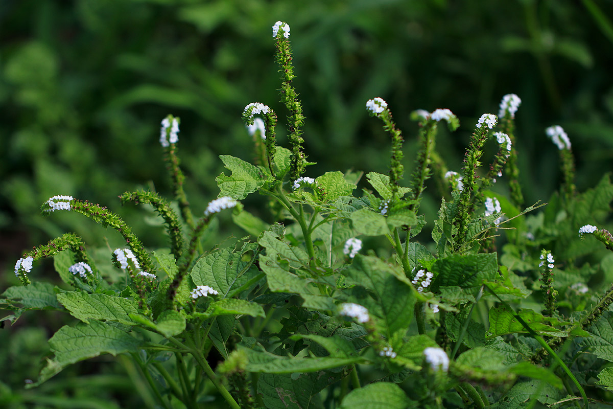
Viny shrub or tall herb, upto 1.5 m high, scarcely woody, bushy, with very dense growth
Shallow tap roots, but occasionally stilt roots are conspicuous
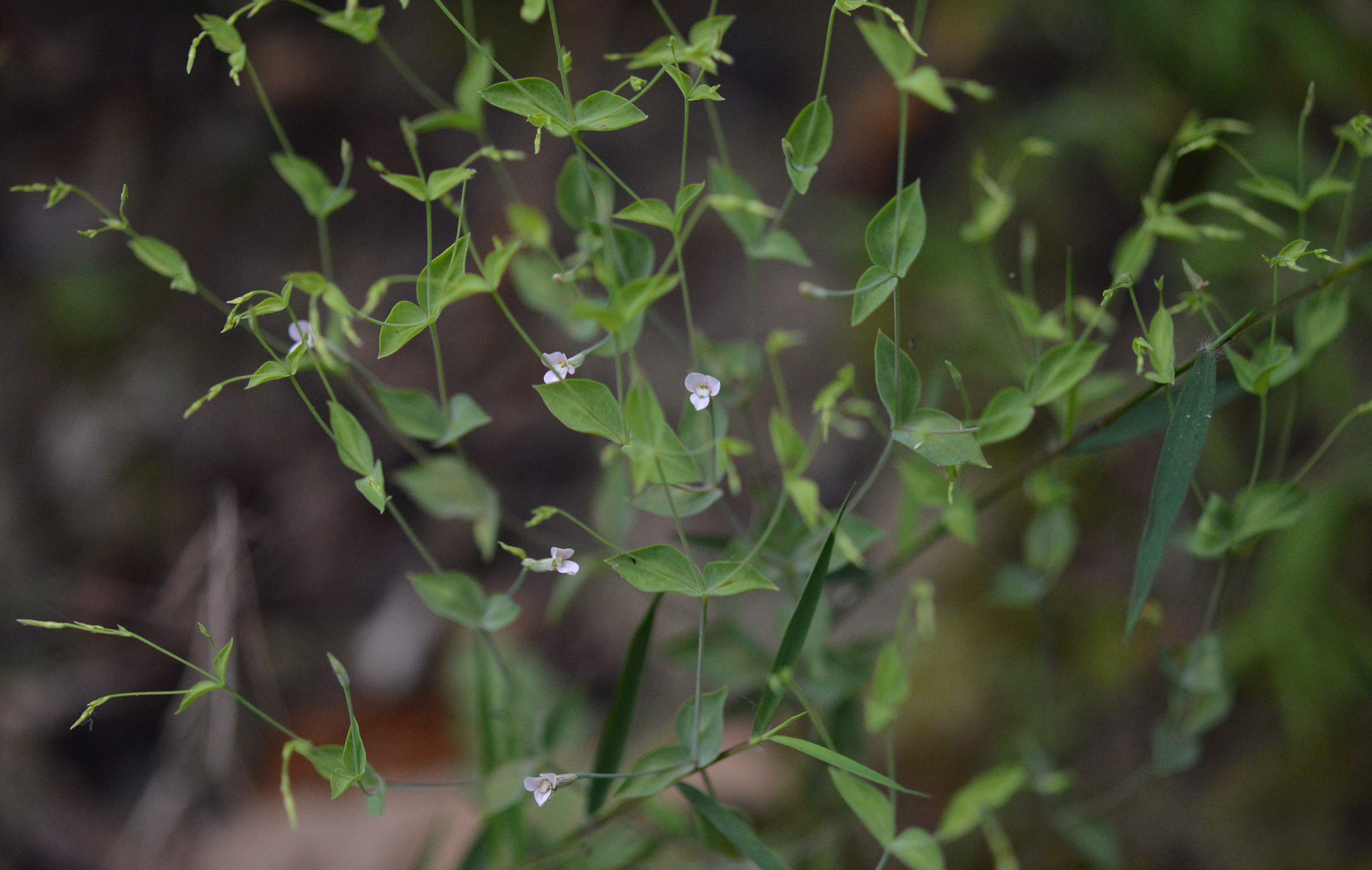
Leaf simple, opposite, decussate, cauline, exstipulate, petiole short, flattened, glabrous, pulvinous to sheathing base
Lamina oblong, elliptic-oblong, spiny margin and wavy, apex acute, basal angled, dorsiventral, green
Inflorescence raceme, spike, upto 16 cm long, peduncled
Flowers bracteate, curved, ovoid, entire, acute, basal cup-shaped, glabrous, greenish, two bracteoles partly covered by bracts
Flower bisexual, typically zygomorphic, complete, erect, sessile, hypogynous,
Sepals 5, polysepalous, twisted, borad, ovate, margin folded, acute, basal curved, coriaceous, greenish , persistent
Petals 1, large, showy, violet, ovate, entire, obtuse, coriaceous, slightly fleshy, slightly curved, margin outerly folded, deciduous
Stamens 4, free filament, erect
Carpels 2, syncarpous, ovary dumbel-shaped, glabrous, terete, ovary superior, two chambered, two ovules in each chamber, axile placentation
Styel 1, terminal, persistent, terete, glabrous
Stigma two bifid.
Fruit 1 cm green and 2.5 - 2.0 cm long, kidney shaped 4 seed drupe, Seed 0.5 - 1.0 cm long
Histology:
Anatomy of leaves described as dorsiventral, leaves showing adaxial and abaxial barrel shaped epidermal cells with thick cuticle. Salt glands present in adaxial side. Each gland have stalk cell and head comprising of variable number of cells. Median vein showed numerous bundles with irregular nature. The whole bundle do not shows a continuous sheath of sclerenchyma. Median vein somewhat paw shaped consists of wide parenchymatous central portion. The lateral vein bundles were somewhat near to the median bundle showing continuous layer of sclerenchyma sheath. The hypodermis is composed of two layers of colourless, polygonal shaped cells below the adaxial epidermal cells. The stomata were confined on abaxial side only. The palisade tissue was two layers in thickness. Vascular bundles collateral. Micro anatomy showed ridges and furrow like appearance in the outer surface. Epidermal cells were straight, glandular trichomes and stomata intermittently seen in the abaxial region. Stomata deeply sunken. Anatomy of stem with one layered thick epidermis. Cortex multilayered with compactly arranged parenchymatous cells. Outer cortex with collenchymas cells and inner cortex consist of parenchyma with air cavities. Growth rings are indistinct therefore classified under diffuse porous wood. Vessels are small to medium sized, even very numerous solitary and round to angular in outline. Vessel lines are indistinct. Vessel cylindrical in nature with alternate inter- vessel pitting and simple perforation plate. Rays were biseriate, homogenous and moderately broad to fine. Xylem showed spiral and annular, helical thickening. Solitary sclereids scattered in the cortex that was seen around phloem cells. Central parenchymatous pith with sclereids and tannin cells. Pith was very large with compactly arranged parenchymatous cells.
Transverse section of fruit anatomy outer cuticle outer cortex consists of sclerieds followed by chlorophyllated polygonal shaped parenchyma cells. Inner portion consists of sclerieds with a groove were present in centre. Tracheids present in inner cortex.
- » Classification and names of artagala
- » Synonyms and definitions of artagala
- » Drug Properties of artagala
- » Chemical Constituents of artagala
- » Standardization of artagala
- » Parts used and Dosage of artagala
- » Morphology and Histology of artagala
- » Distribution and Conservation of artagala
- » Cultivation of artagala
- » artagala in the market
- » Medicinal Uses of artagala
- » Researches and clinical trails of artagala
- » artagala in other sytems of medicine
- » Ayurvedic formulations with artagala
- » Images of artagala
Website Administrator
This article is incomplete. If you feel you can make a contribution, please let us know by sending email to mail@ayushvedah.com, we may consider your request. Thank you.