kasamarda :

Morphology:
Cassia occidentalis, is a much branched, smooth, half woody herb or shrub about 0.8 to 1.8 m tall.
- Stem is erect, and without hairs.
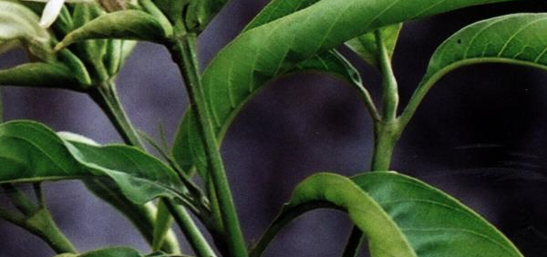
- Leaves are lanceolate or ovate-lanceolate, bipinnately compound, and about 20 to 25 cm in length. Each pinna has four to seven pairs of leaflets, which are 3 to 9 cm in length, and 2 to 4 cm in width, and arranged oppositely. Leaflets are ovate or ovate lanceolate in shape with a long, fine pointed tip. Each leaf has a distinct spherical shaped gland, which is located about 0.3 to 0.5 cm from the base of the petiole.
- Inflorescence is a terminal or axillary raceme. Flowers are yellow colored, and about 2 cm long, and 3 to 4 cm wide.
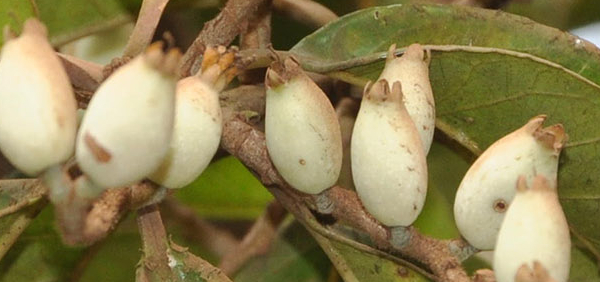
- Fruit is a pod / legume, compressed, 8 to 12 cm long, 0.7 to 1 cm wide, and curved slightly upwards. Each pod contains 20 to 30 seeds, which are ovoid in shape, smooth, shiny, and dull brown to dark olive-green in colour.
Histology:
Transverse section of the leaf containing midrib region.
Epidermis :- Both the surfaces are covered by upper and lower epidermal layers. Both the upper and lower epidermis possesses a prominent cuticle and sunken stomatas (Pandey, 1969). Both the upper and lower epidermis possesses trichomes and hairs. The hairs are non-glandular, unicellular, conical, often curved (Pandey, 1970). Trichomes are glandular, multiseriate slatk, multi cellular and with capitate head Shah and Gopal (1971). Stomatas in cassia occidentalis are intermed between diacytic and paracytic type. According to Metcalfe and chalk, 1950 the family Caesalpiniaceae have paracytic and anomocytic stomatas.
Mesophyll :- The tissue of the leaf that lies between the upper and the lower epidermis and between the vein consists of typically thin walled parenchymatous cells called mesophyll. This mesophyll consist of palisade and spongy parenchyma. The mesophyll cells are loosely arranged, parenchymatous in nature and contain rosette or prismatic crystals of calcium oxalate. The palisade tissue is exhibited both near the upper and the lower epidermis and they consist of a single layer of elongated narrow columnar cells containing chloroplastids. The vascular bundles are embedded in this region.
Vascular Bundles :- A transverse section through the midrib region shows a single arc shaped vascular bundle ensheathed by sclerenchymatous cells. The vascular bundle is collateral with xylem on the upper region (towards upper epidermis) while the phloem is in the region towards the lower epidermis. Xylem is in the center and is surrounded by phloem, protoxylem elements are towards upper epidermis. Vascular bundle is collateral and open type. This group of bundle is protected both above and below by an arc of lignified fibers, which is somewhat ovate in shape above and crescent shaped below. This arc of fiber have on their outer surface a layer of cells most of which contain prism of calcium oxalate.
Anatomy of Root Root is diarch with typical dicotylednous secondary growth. The dicotyledonous root possess a limited number of radial Vascular bundles with exarch Xylem.
Transverse Section of the Root Periderm :- Outermost layer is epiblema or piliferous layer which due to secondary growth is replaced by cork.. Cortex :- 1. It consist of thin walled parenchyma with numerous intercellular spaces. 2. The cell possesses large number of chloroplasts. After the formation of periderm, cortex and pericycle are peeled off.
Endodermis :-Cortex is followed by well demarcated and single layered Endodermis. But due to development of periderm in the pericycle, endodermis is sloughed off and and is therefore not visible in the old structure. Pericycle :- It follows endodermis and it is single layered. It is the place where cork cambium originated to four periderm.
Vascular tissue system :-
1. The vascular bundles are radial and exarch.
. Xylem and phloem form equal numbers of seperate bundles with protoxylem towards the pericycle (Exarch).
3. It consist of primary phloem, secondary phloem cambium, secondary xylem, medullary rays and primary xylem.
4. The secondary Vascular tissue form a continous cylinder just below the pericycle.
5. The Pericycle region contains a ring of crushed and obliterated groups of primary phloem fibres or the primary phloem is present in the form of patches.
6. Secondary phloem group that occur below patches of primary phloem are massive.
7. Primary xylem groups are located close to the center of axis. Protoxylem are directed away from the center (condition exarch).
8. Cambium is present between secondary phloem and secondary xylem which can be unistratose to multistratose.
9. Secondary xylem lies below the cambium. It is divided into many smaller and larger regions due to wide medullary rays which pass through it.
10. Phloem consist of sieve tubes, companion cells and phloem parenchyma.
11. Xylem consist of Vessels, tracheilds and thick walled xylem parenchyma.
Pith :- Pith is very small present in the center of the axis or altogether absent .
Anatomy of Stem Transverse section of stem Epidermis :-
1. It is the outermost single layer and consist of stomatas and trichomes.
2. Outer walls of the cells are greatly thickened and heavily cutinized.
3. Cell of epidermis are almost rectangular and do not possess inter cellular spaces.
Cortex :-
1. The region lying next to the epidermis is the cortex.
2. Cortex is few layered deep and is Chlorenchymatous in nature.
Endodermis :- This is single layer which separates the cortex from the vascular bundles. The cells lack Casparian strips but contain starch. Hence, called starch sheath. Cells are barrel-shaped, elongated, compact and having no intercellular spaces among them.
Pericycle :-
1. It follows endodermis and is sclerenchymatous in nature.
2. Sclerenchymatous patches are present over phloem groups of Vascular Bundles. These are called Hard Bast.
Vascular Bundles :-
1. These are present in a ring.
2. Each vascular bundle is conjoint, collateral, endarch and open type.
3. Xylem which is present near the center of the stem is known as protoxylem and the xylem towards the peripheral part of the stem is Metaxylem. This condition is endarch. .
4. It consist of primary xylem, secondary xylem, cambium, primary phloem, secondary phloem medullary rays or vascular rays.
5. Xylem consist of vessels, tracheids, xylem parenchyma and fibres.
6. Phloem consists of sieve tubes, companion cells and phloem parenchyma.
7. A few layers of cambium are present between xylem and phloem elements.
8. Cambium divides towards outside to form secondary phloem and after the formation of secondary phloem the primary phloem becomes crushed and function less.
9. Phloem rays are formed in the vascular tissue developed by the cambium.
10. Cambium divides on its inner side to form secondary xylem
11. Xylem rays are formed radially in the secondary xylem. They are strap or ribbon like. They run as a continous band to the secondary phloem, thus rays are called medullary rays or pith rays or vascular rays.
Pith :- The central part of the section is occupied by paremchymatous pith with numerous tannin cells, sphaero-crystals and starch grains. The paremchymatous cells have distinct intercellular spaces.


