shleshmataka :

Morphology:
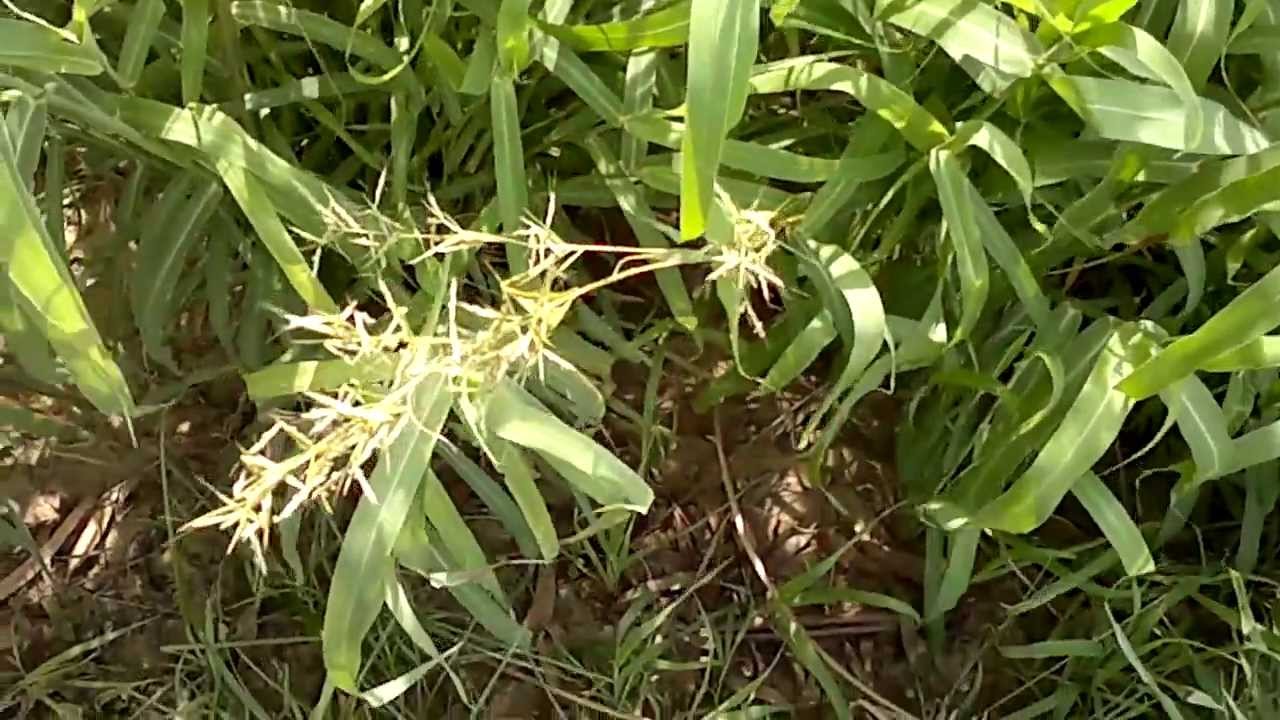
Root - 5-12 cm long, 1-7 mm thick, oblique and gradually tapering, bearing a few rootlets; external surface, dirty brown; fracture, short. Stem - Glabrous, cylindrical, hairy, slightly branched; 10-17 cm long, 1-8 mm thick, grooved and ribbed; basal region of branches greenish-brown, apical region dark green, bearing a number of flowers; fracture, short. Leaf - Simple, dark-green, smooth, alternate, opposite, exstipulate, 2.5-5 cm long, 1.8-3.6 cm broad, elliptical, lanceolate, obtuse or acutely toothed; shape and size variable; petiole short; odour, slightly characteristic.Histology:
Root - Mature root shows 4-5 layered cork, consisting of tabular, tangentially elongated, thick-walled cells filled with reddish-brown contents; secondary cortex consists of a wide zone of thin-walled, parenchymatous cells having a few resin ducts; secondary phloem, a narrow zone, composed of sieve elements and phloem parenchyma, traversed by phloem rays; xylem well-developed, composed of vessels, tracheids, fibres and xylem parenchyma, traversed by 1-5 seriate xylem rays; xylem vessels usually solitary or 2-4 in groups with reticulate thickening; fibres aseptate and pointed. Stem - Mature stem shows several bulges at places and consists of a single layered epidermis, externally covered with a striated cuticle; a number of epidermal cells elongate to form multicellular covering and T-shaped trichomes with 2-6 celled stalk; cortex 3-5 layers of thin-walled, tangentially elongated parenchymatous cells, a few filled with reddishbrown content, bulges show a few layers of collenchyma between epidermis and parenzhymatous cortex; endodermis single layered, composed of barrelshaped cells; pericycle occurs in the form of groups of pericyclic fibres; phloem consists of strands of sieve tubes, companion cells and phloem parenchyma; xylem consists of vessel, parenchyma and fibres; xylem vessls show reticulate thickening; parenchyma in abundance and paratracheal; fibres thick-walled, aseptate, short, with pointed ends; medullary rays 2-11 cells wide; central portion occupied by pith composed of hexagonal to polygonal, thin-walled parenchymatous cells; a few simple starch grains present in cortical cells; cluster crystals of calcium oxalate occasionally found in pith. Leaf - Petiole - shows a somewhat circular outline with two lateral projections one on each side; epidermis on both surfaces, covered externally with striated cuticle and have both type of trichomes as described in case of stem, followed by 2-3 layers of collenchyma on upper and lower side; stele composed of three collateral vascular bundles located in centre, central one larger and lateral two smaller; ground tissue composed of thin-walled parenchymatous cells, a few having oil globules and rosette crystals of calcium oxalate. Midrib - shows similar structure as described in petiole except for 1 or 2 layers of collenchymatous cells below both epidermis and a single vascular bundle in centre; oil globules and rosette crystals of calcium oxalate present in a few cells of ground tissue. Lamina - shows dorsiventral structure; epidermis single layered on either surface, composed of thin-walled, tangentially elongated cells, covered externally with striated cuticle; trichomes similar to those of stem; palisade single layered; spongy parenchyma 4-5 layered, loosely arranged cells; vascular bundles embedded in spongy parenchyma; rosette crystals of calcium oxalate and oil globules present in this region; anomocytic stomata present on both surfaces.- » Classification and names of shleshmataka
- » Synonyms and definitions of shleshmataka
- » Drug Properties of shleshmataka
- » Chemical Constituents of shleshmataka
- » Standardization of shleshmataka
- » Parts used and Dosage of shleshmataka
- » Morphology and Histology of shleshmataka
- » Distribution and Conservation of shleshmataka
- » Cultivation of shleshmataka
- » shleshmataka in the market
- » Medicinal Uses of shleshmataka
- » Researches and clinical trails of shleshmataka
- » shleshmataka in other sytems of medicine
- » Ayurvedic formulations with shleshmataka
- » Images of shleshmataka

Dr Mohamed Rifas
A dual country licensed Ayurveda Physician, currently working in Dubai as Head of Ayurveda and Yoga department. He also works as visiting doctor for Dr Hassan Ayurveda Brain and Spine Specialty Hospital. He is specialized in joint issues, brain and spine related issues and gastrointestinal issues with respect to anxiety.
Doctor profile: Click here